Cells Intro
There is a pretty good interactive site here. The left panel gives you the basics of cells. We may use some of the other animations later. It is not required that you go to and use this site. But, you may find it helpful.
It will cover most of the reading.
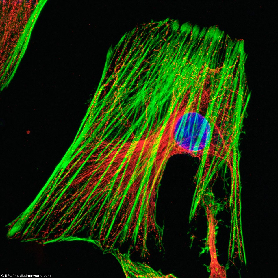
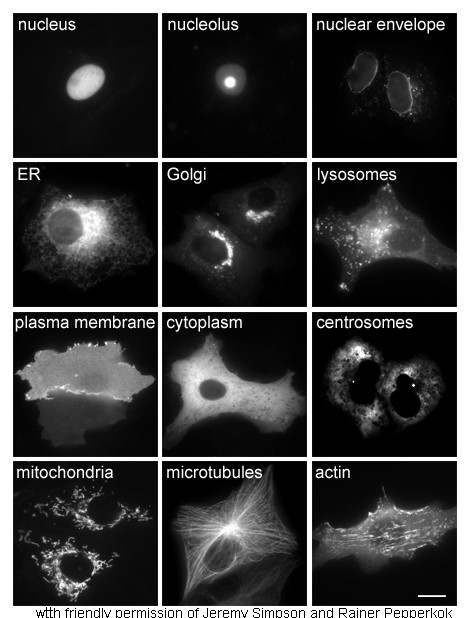
Below I’m including some real micrographs of what the components of cells look like. I can explain the technique in great detail later, if you like. But, for now, just know that we have tools that allow us to make any structure we want “fluoresce” or “glow” in a special microscope. These are not the best images. But you get the idea.
It will cover most of the reading.
All living things are made of cells.
- Why? Because I said so. Well, not because I said so. This is something called the “cell theory of life” and it builds into our definition of life the requirement for cells. There are a lot of ways in which this makes sense. However, there are many biological entities, most notably, viruses, which are not cellular and at least fulfill many of the other requirements for being alive.
Components of a cell
- I want you to know the names and general descriptions of the major organelles.
- Nucleus: large structure that houses the chromosomes (DNA). All RNA for the cell is made there, using the DNA as a template. It is encased by a double membrane. That is, two complete lipid bilayers. There are pores, or holes, for the transport of stuff out and in.
- Nucleolus: dense structure in the nucleus. It is the site of ribosome assembly. Ribosomes are then transported to the cytoplasm through the pores. Some scary numbers: a single mammalian cell may have 10,000,000 ribosomes and about 10,000,000,000 protein molecules (numbers from British Society for Cell Biology).
- Ribosomes are RNA/Protein machines that synthesized proteins. They show up as little black dots in electron micrographs either stuck to rough ER or free.
- Rough endoplasmic reticulum: Stack of membrane tubes near the nucleus. Site of synthesis of proteins that either end up in membranes or are secreted out of the cell (secreted merely means transported out of the cell). They are “rough” because they are dotted with ribosomes. Again, there are “Free” ribosomes that are not attached to the ER that synthesize proteins not bound for export or other membrane compartments.
- Smooth endoplasmic reticulum: Lacks ribosomes. This is the name given to stripy membrane tubes that have many varied functions depending on cells. In all cells, it is the site of phospholipid synthesis. In addition, in specialized cells, it is the site of steroid synthesis or used to manage calcium ion concentrations in cells that really use a lot of that. In liver cells, the break down of various toxins by enzymes takes place here. In the electric organs of fish, they stack up as battery cells using ion pumps to establish voltage.
- Golgi apparatus: Stacks of membranes that are between the rough ER and the plasma membrane. Proteins are transported from the ER to the Golgi in spheres of membrane called “vesicles.” In the Golgi, proteins are processed (usually meaning sugar molecules are added to specific amino acid residues).
- Plasma membrane or cell membrane: the lipid bilayer that keeps the outside out and the inside in. It also has many proteins in it, which may function, for example, as channels allowing transport of nutrients, or allow cells to grab other surfaces or detect the presence of various molecules: signals to change behavior, or example, or simply to hold on to other cells.
- Endocytic Vesicles: Spherical membranes that derive from the plasma membrane. Imagine them as pinching off from the membrane and taking in with the membrane any proteins in the area that pinches off. They are sites for “recycling” components of proteins. Viruses often exploit this as a way into cells.
- Lysosome: the next step for many of the endocytic vesicles. Highly acidic vesicles with digestive enzymes that fuse with the endocytic vesicles to allow the break-down of their contents. In fungi and plants, many scientists (and your book) say “lysosomes” don’t exist per se. Instead, larger membrane structures called “Lytic vacuoles” carry out the same process. What’s in a name?
- All together, numbers 3-9 (actually starting at the nuclear envelope) make up sort of one continuous super-organelle comprising a system of membranes. If I were to make radioactive lipids and put them in the cell, so I could detect the radiation where ever it was, I would find the “hot” lipids show up first in the smooth ER and nuclear envelope, and then later would be detected in each of the other places as time passed, in the order I presented.
- Mitochondria: these are the sites in which the high energy molecules from your food are broken down to low energy products (water and CO2) and the released free energy is captured in other high-energy molecules such as ATP, used to help run many reactions in your body. Mitochondria have their own chromosome (circular, like bacteria), their own ribosomes and their own tRNA. The function of all these components resembles that of bacteria. It is thought that mitochondria derived evolutionarily from bacterial cells that became part of a larger cell.
- Cytoskeleton: worst representation ever in the diagrams. We will look more at this later. It is a network of fibers made of the proteins actin (microfilaments), tubulin (microtubules) and Keratin (intermediate filaments…actually, there are many forms of each of these proteins). Microfilaments and microtubules are constantly changing. Microfilaments are usually involved when cells are migrating, are found more at the edges of the cell and at points where cells are attached to surfaces. Microtubules all grow out of centrioles or similar structures (generically known as “microtubule organizing centers” or MTOC). Again, somewhat semantically, higher plants don’t have well defined centrioles, but do have a “centrosome” as the MTOC. In a generic animal cell, the MTOC usually is the centriole, near the nucleus. However, the tail of sperm and many other things that stick out of cells and allow them to move (wag like the tail of sperm…called either “flagella” or “cilia” are made of microtubules and they each require their own set of centriole-like structures).
- Intermediate filaments are more permanent and are used to link cells together in very strong tissues (like skin). You only find them in cells that aren’t going any where anytime soon.
Unique to plants:
- Cell wall: cellulose-based structure that provides rigid “box” around cell.
- Vacuole (or central vacuole): large, fluid-filled region of the cell, surrounded by membrane. Used to maintain pressure in the cell, keeping the cell pressed out against the cell wall.
- Chloroplast: site of photosynthesis. Like the mitochondria, these have their own DNA, ribosomes and tRNA. They also appear to have been bacterial in origin…long ago.
- “Higher” plants (all the plants you know well…except maybe kelp), don’t have true centrioles. This is really a distinction without a difference. They have MTOCs, but lack the clear cylindrical tubes that we call centrioles. Kelp, which is not considered a higher plant, has a motile single-cell phase that swims using flagella. These have centrioles associated with them.
Below I’m including some real micrographs of what the components of cells look like. I can explain the technique in great detail later, if you like. But, for now, just know that we have tools that allow us to make any structure we want “fluoresce” or “glow” in a special microscope. These are not the best images. But you get the idea.